- Received May 10, 2022
- Accepted May 27, 2022
- Publication June 22, 2022
- Visibility 3 Views
- Downloads 0 Downloads
- DOI 10.18231/j.jooo.2022.017
-
CrossMark

- Citation
Prodigious presentation of oral squamous cell carcinoma: early diagnosis and treatment – A case report
Introduction
Squamous cell carcinoma is one of the most trivial forms of cancer of the head and neck region.[1] Its prevalence is different in various parts of the world; in unindustrialized countries, like India, it is most commonly diagnosed in male patients whereas, in the Western world, it is responsible for 1–4% of all cancers.[2] Head and neck cancer is fourteenth in terms of incidence but thirteenth in terms of mortality among all the other cancers.[3] More than 90% of cancer cases in head and neck regions are OSCCs.[4]
The most common predisposing factors are tobacco consumption (including betel quid, tobacco with lime, beedi, and hookah smoking),[5] alcohol, ultraviolet radiation (specifically for lip cancer), but several other factors such as human papillomavirus (HPV) and Candida infections, nutritional deficiencies have been also associated[6] especially in the Western world[7] whereas consumption of smokeless tobacco and areca nut products are the main etiological factors associated with OSCC in South Asian countries.[8] Its etiology may also be related to gene mutations; however, no specific gene has been identified to date.[9] Environmental factors such as smoking, irradiation, and viral infection may increase the risk of oral and oropharynx OSCC by activation of proto-oncogenes (ras, myc, EGFR) or inhibition of tumor suppressor genes (TB53, pRb, p16).[10]
Most of the oral and oropharynx OSCC cases occur in elderly male patients, with stratified squamous epithelial lining of the Buccal mucosa, tongue, floor of the mouth, palate, lip, tonsils, and tongue is the most commonly affected sites.[11] It presents as an ulcerated lesion with a central necrotic area and rolled up margin.[12], [13]
Many cases remain undiagnosed until the advanced stage when metastases have happened.[14] Even for the OSCC patient who receives surgical resection, local recurrence is another cause for treatment failure.[15] Therefore, a highly sensitive and specific screening approach is imperatively needed for diagnosis and prognosis. Currently, the visual inspection of the mouth combined with histopathology is still the gold standard method for oral cancer screening.[16], [17]
Early detection of neoplasms is indispensable to reduce morbidity and mortality associated with oral cancers. Dentists play a leading role in the early detection of oral cancers, thus increasing the survival rates of patients.
Presentation of Case
A 57-year-old male patient came to the Department of Oral Medicine and Radiology with the chief complaint of an ulcer in the right cheek region for 1 month. The patient gave a history of smoking beedi 8-10 per day for the last 15 years. He stopped smoking from the last 3 months. Medical history and family history were non-contributory. On extraoral examination single right submandibular lymph node was firm in consistency, tender, and mobile on palpation. No extraoral swelling was evident. ([Figure 1])

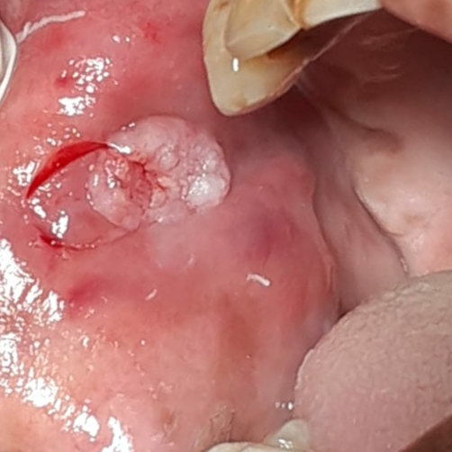
On clinical examination right buccal mucosa reveals a solitary well-defined proliferative growth of approximately 1.68x1.88cm in greatest dimension, sessile in nature, of normal mucosal color extending 3.5cm posterior to the retrocommisural area at the level of the occlusal plane. ([Figure 2])

The surface appeared irregular, lobulated with rolled-out edges. It was roughly ovoid in shape. On palpation, the growth was non-tender, not fixed to deeper structures, firm in consistency with mild-indurated margins. It had a rough surface and did not bleed on manipulation. The single right submandibular lymph node was palpable.
Clinical diagnosis
Malignancy in right buccal mucosa.
Differential diagnosis
Traumatic fibroma
Reactive hyperplasia
Squamous papilloma
Warts
Keratoacanthoma
Investigations
Patient was advised routine blood investigations – complete blood count (CBC), random blood sugar (RBS), bleeding time (BT), clotting time (CT) and COVID test.
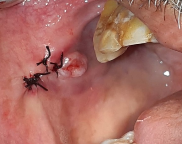
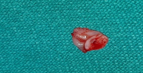
All the reports were within normal limits. Hence, the patient was advised incisional biopsy to confirm the diagnosis. ([Figure 3], [Figure 4])
The specimen was sent for histopathological examination. ([Figure 5])
Histipathological features
Epithelium is dysplastic stratified squamous and exhibits tumor islands invading the connective tissue stroma. The tumor islands exhibit individual cell keratinization, altered nuclear cytoplasmic ratio, mitotic figures and keratin pearl formation. The surrounding connective tissue is fibrovascular and shows dense chronic inflammatory cell infiltrate ([Figure 6]) All these features are suggestive of Well Differentiated Squamous Cell Carcinoma.
The treatment includes wide excision of the lesion including 1mm of normal marginal tissue followed by intraoral suturing by the department of Oral Surgery and anesthesia.

Discussion
Squamous cell carcinoma is defined as “a malignant epithelial neoplasm exhibiting squamous differentiation as characterized by the formation of keratin and/or the presence of intercellular bridges”. (Pindborg JJ et al, 1997).
Squamous cell carcinoma also called as the epidermoid carcinoma is the most common malignant neoplasm of the oral cavity. Although any intraoral site may be involved but certain sites are more periodically involved than others. A number of rare conditions predispose to the development of oral cancer, such as xeroderma pigmentosum, Fanconi’s anemia, and Bloom’s syndrome. Classic microscopic histopathologic alterations observed with squamous cell carcinoma include:
Enlarged nuclei as well as cell size
Large and prominent nucleoli
Increased nuclear/cytoplasmic ratio
Hyperchromatic (dark staining nuclei
Dyskeratosis (premature keratinization of cells
Increased and/or aberrant mitotic activity
Different regions have different addictive habits associated with OSCC therefore, the sites involved in tumor initiation also are different.
Conclusion
Squamous cell carcinoma is the most common malignant epithelial neoplasm with varied oral presentations. Therefore, the dentist should be aware of the characteristics of the disease. Distant metastasis is the most inevitable complication as the disease progresses. Hence, it is important to diagnose correctly and timely as there are more chances for misdiagnosis because its clinical presentation can mimic inflammatory gingival lesions.
Source of Funding
None.
Conflict of Interest
The authors declare no conflict of interest.
References
- L Capparuccia, L Tamagnone. Semaphorin signaling in cancer cells and in cells of the tumor microenvironment--two sides of a coin. J Cell Sci 2009. [Google Scholar]
- P Joshi, S Dutta, P Chaturvedi. Head and neck cancers in developing countries. Rambam Maimonides Med J 2014. [Google Scholar]
- A Al-Jaber, L Al-Nasser, A El-Metwally. Epidemiology of oral cancer in Arab countries. Saudi Med J 2016. [Google Scholar]
- P Tandon, A Dadhich, H Saluja, S Bawane, S Sachdeva. The prevalence of squamous cell carcinoma in different sites of oral cavity at our Rural Health Care Centre in Loni, Maharashtra - a retrospective 10-year study. Contemp Oncol (Pozn) 2017. [Google Scholar]
- MP Singh, V Kumar, A Agarwal, R Kumar, ML Bhatt, S Misra. Clinico-epidemiological study of oral squamous cell carcinoma: A tertiary care centre study in North India. J Oral Biol Craniofac Res 2016. [Google Scholar]
- NW Johnson, P Jayasekara, AAHK Amarasinghe. Squamous cell carcinoma and precursor lesions of the oral cavity: epidemiology and etiology. Periodontol 2000 2011. [Google Scholar]
- S Graham, H Dayal, T Rohrer. Dentition, diet, tobacco, and alcohol in the epidemiology of oral cancer. J Natl Cancer Inst 1977. [Google Scholar]
- SS Muttagi, P Chaturvedi, R Gaikwad, B Singh, P Pawar. Head and neck squamous cell carcinoma in chronic areca nut chewing Indian women: Case series and review of literature. Indian J Med Paediatr Oncol 2012. [Google Scholar]
- A Krishna, S Singh, V Kumar, US Pal. Molecular concept in human oral cancer. Natl J Maxillofac Surg 2015. [Google Scholar]
- BW Neville, DD Damm, CM Allen, JE Bouquot. . Oral and maxillofacial pathology 2009. [Google Scholar]
- DJ Weatherspoon, A Chattopadhyay, S Boroumand, I Garcia. Oral cavity and oropharyngeal cancer incidence trends and disparities in the United States: 2000-2010. Cancer Epidemiol 2015. [Google Scholar]
- FR Pires, AB Ramos, JBC Oliveira, AS Tavares, PSR Luz, TCRB Santos. Oral squamous cell carcinoma: Clinicopathological features from 346 cases from a single oral pathology service during an 8-year period. J Appl Oral Sci 2013. [Google Scholar]
- BW Neville, TA Day. Oral cancer and precancerous lesions. CA Cancer J Clin 2002. [Google Scholar]
- YS Cheng, T Rees, J Wright. A review of research on salivary biomarkers for oral cancer detection. Clin Transl Med 2014. [Google Scholar]
- JT Chen. Glycoprotein B7-H3 overexpression and aberrant glycosylation in oral cancer and immune response. Proc Natl Acad Sci U S A 2015. [Google Scholar]
- LH Hartwell. Reply to Galvão-Moreira and da Cruz: Saliva biomarkers to complement the visualization-based oral cancer detection. Proc Natl Acad Sci U S A 2017. [Google Scholar]
- LV Galvão-Moreira, MC DaCruz. Saliva protein biomarkers and oral squamous cell carcinoma. Proc Natl Acad Sci U.S.A 2017. [Google Scholar]