- Visibility 205 Views
- Downloads 29 Downloads
- DOI 10.18231/j.jooo.2020.044
-
CrossMark
- Citation
A rare case of lipoma on the buccal mucosa: A case report
- Author Details:
-
Shaul Hameed K *
-
Imran Mohtesham
-
Yasir Alyahya
-
Anoop Kurian Mathew
-
Mariyam Nishana M
Introduction
The lipoma is listed as a common tumor of mesenchymal origin seen in areas where fat is located in the body. They are usually considered as benign soft tissue tumors comprising of mature adipocytes histopathology, the cells of the lipoma differ metabolically from normal fat cells even though they are histologically similar.[1] Subcutaneous occurrence is more often than in deeper tissues.[2] The etiology of lipoma is unknown. They are known to develop in and around shoulders, trunk, axilla, and neck. Oral lipoma accounts for 4.4% of oral soft tissue benign tumors.[3] Clinically they are usually manifested as asymptomatic, yellowish in color, soft doughy consistency swelling. The usual site of oral occurrence is buccal mucosa, tongue, and floor of the mouth. Fourth and fifth decades of life in males predominantly known to have a lipoma. Occurrence on lips, palate, vestibule, and salivary gland may interfere with mastication and speech.[4] We hereby present a case report of 52 years old male from south India with a lipoma on the buccal mucosa diagnosed and treated with surgical excision and confirmed by histopathological diagnosis.
Case Report
A 52-year-old male patient presented to the Department of Oral Medicine and Radiology for the management of swelling at the left buccal mucosa. The swelling was small in size initially and increased in size gradually over the past five years. There was no history of any associated symptoms or bleeding/discharge from swelling. On local examination, it was 2 cm x 2 cm in diameter with pedunculated base yellowish in color. The surface appeared to be normal, on palpation. The swelling was doughy inconsistency, slippery, and not tender. [[Figure 1], [Figure 2]]
Based on the clinical examination and chair side investigation provisional diagnosis of lipoma was made. An excisional biopsy was done. The tissue was sent for histopathological diagnosis.
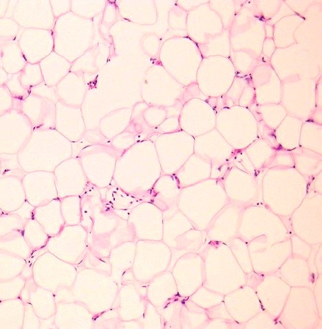
Routing H and E staining were done. On histopathological examination of the lesion, it depicted mature adipose tissue admixed with collagen streaks with well-demarcated surrounding connective tissues. The thin fibrous capsule exhibited a lobulated pattern. The section also showed few blood vessels in the connective tissue stroma. [[Figure 3]] Based on the clinical and histopathological findings a diagnosis of lipoma was given. Post-operative follow up revealed uneventful healing with no recurrence.


Discussion
The first oral lipoma was reported in 1848 and was termed as Yellow Epulis.[3] Lipoma has commonly encountered tumor origin of adipose tissue, but very rarely present in the oral cavity with a prevalence of 1in 5000.[5] It can occur at any age but more frequently seen in the male group with age above 40 years. Our case also was a male patient with 52 years of age. The clinically intraoral lipoma is low grade, slow-growing, it takes 10 years for the patient to seek attention from the date of first notice with a mean value of 2 years.[6], [7]
Oral lipomas (OLs) occur most commonly in the parotid region followed by the buccal mucosa, tongue, floor of the mouth, and palate.[8] When present in the buccal mucosa it will be of round or oval with well-defined mass. Usually rigid and in some cases it is soft depends on the fibrous content. In our case, it was round well defined and rigid. It is a simple criterion to diagnose clinically based on the appearance unless located deeply. Clinically, the lipoma is described as a slow-growing, well-circumscribed mass of the soft tissue.[9]
The histopathological differential diagnosis for lipoma is leiomyoma, fibroma, pleomorphic adenoma of the minor salivary gland, epidermoid cyst, and papilloma.[10] Histopathological there will be a nodular structure with connective tissue covered by stratified squamous epithelium. Lipoma rather than liposarcoma is diagnosed if the lesion does not show any of the following: lipoblastic proliferation, variation in adipocyte size, atypical and enlarged adipocyte nuclei, hyperchromatic, or bizarre stromal cells in fibrous septa, between adipocytes or in vessel walls.[8]
In leiomyoma's, there will be bundles of interlacing spindle-shaped smooth muscle cells, elongated nuclei blunt-ended, and pale staining. Pleomorphic adenomas will be encapsulated, well-circumscribed and the capsule may show tumor cell infiltrate.
Papilloma's contains keratinized squamous cell proliferation in a finger-like pattern with fibrovascular connective tissue.[11]
In the present case, it showed classic features of adipose tissue with blood vessels and an 8-10 cell layer of atrophic stratified squamous epithelium strongly suggesting Lipoma on the buccal mucosa. Histopathological confirmation plays a major role in these cases because the treatment differs for if the results come out to be different lesions. Although the growth of oral lipomas is usually limited, they can reach a great dimension, interfering with speech and mastication.[12] The main treatment of lipoma is complete excision of the mass which was done for the present case. However, recurrence has not been reported so far after adequate excision.[13]
Conclusion
Lipoma when present in the oral cavity is low grade, slow-progressing asymptomatic unless the mass causes interaction with the mastication and aesthetics issues. It develops subcutaneously and fewer times involves deeper tissues. Complete surgical excision is the best way to prevent a recurrence.
Source of Funding
None.
Conflict of Interest
The author(s) declare(s) that there is no conflict of interest regarding the publication of this article.
References
- J L Gall, S Laurence, B Khalifa, Ce B Rombi, C Mauprivez, B Lefèvre. Intraoral fibrolipoma: case report and review of literature. J Oral Med Oral Surg 2018. [Google Scholar] [Crossref]
- L P Zhong, S F Zhao, G F Chen, F Y Ping. Ultrasonographic appearance of lipoma in the oral and maxillofacial region. Oral Surg Oral Med Oral Pathol Oral Radiol Endod 2004. [Google Scholar]
- E C Studart-Soares, F W Costa, F Sousa, A P Alves, R L Osterne. Oral lipomas in a Brazilian population: A 10-year study and analysis of 450 cases reported in the literature. Med Oral Patol Oral Cir Bucal 2010. [Google Scholar] [Crossref]
- B S Manjunatha, G S Pateel, V Shah. Oralfibrolipoma-a rare histological entity: Report of 3 cases and review of literature. J Dent 2010. [Google Scholar]
- B W Neville, D D Damm, C M Allen, J E Bouquot. . Oral and Maxillofacial Pathology. Soft tissue tumors 2002. [Google Scholar]
- J C Hatziotis. Lipoma of the oral cavity. Oral Surg, Oral Med, Oral Pathol 1971. [Google Scholar] [Crossref]
- J E Horton. Lipomas of the tongue: report of a fibrolipoma. Oral Surg Oral Med Oral Pathol 1968. [Google Scholar]
- L Bodner, R Shaco-Levy, Intramuscular. Infiltrating) Lipoma of Floor of the Mouth. Case Rep Med 2018. [Google Scholar]
- L Goutzanis, A Chliaoutakis, D Kalyvas. Bilateral buccal space lipoma: A rare case presentation. J Clin Exp Dent 2019. [Google Scholar] [Crossref]
- R O Greer, J F Richardson. The nature of lipomas and their significance in the oral cavity. Oral Surg, Oral Med, Oral Pathol 1973. [Google Scholar] [Crossref]
- B W Neville. . Clinical review of oral and maxillofacial surgery 2008. [Google Scholar]
- R L V Osterne, R M B Lima-Verde, E Turatti, C F W Nonaka, R B Cavalcante. Oral cavity lipoma: a study of 101 cases in a Brazilian population. J Bras Pathol Med Lab 2019. [Google Scholar] [Crossref]
- N Dehghani, F Razmara, T Padeganeh, X Mahmoudi. Oral lipoma: Case report and review of literature. Clin Case Rep 2019. [Google Scholar] [Crossref]
How to Cite This Article
Vancouver
K SH, Mohtesham I, Alyahya Y, Mathew AK, M MN. A rare case of lipoma on the buccal mucosa: A case report [Internet]. J Oral Med Oral Surg Oral Pathol Oral Radiol. 2025 [cited 2025 Sep 08];6(4):205-207. Available from: https://doi.org/10.18231/j.jooo.2020.044
APA
K, S. H., Mohtesham, I., Alyahya, Y., Mathew, A. K., M, M. N. (2025). A rare case of lipoma on the buccal mucosa: A case report. J Oral Med Oral Surg Oral Pathol Oral Radiol, 6(4), 205-207. https://doi.org/10.18231/j.jooo.2020.044
MLA
K, Shaul Hameed, Mohtesham, Imran, Alyahya, Yasir, Mathew, Anoop Kurian, M, Mariyam Nishana. "A rare case of lipoma on the buccal mucosa: A case report." J Oral Med Oral Surg Oral Pathol Oral Radiol, vol. 6, no. 4, 2025, pp. 205-207. https://doi.org/10.18231/j.jooo.2020.044
Chicago
K, S. H., Mohtesham, I., Alyahya, Y., Mathew, A. K., M, M. N.. "A rare case of lipoma on the buccal mucosa: A case report." J Oral Med Oral Surg Oral Pathol Oral Radiol 6, no. 4 (2025): 205-207. https://doi.org/10.18231/j.jooo.2020.044
