Introduction
According to WHO, histological typing of odontogenic tumors, AOT is defined as ‘A tumor of odontogenic epithelium with duct – like structures and varying degrees of inductive change in the connective tissue. The tumor may be partly cystic, and in some cases the solid lesion may be present only as masses in the wall of a large cyst.1 Earlier it was known as adenoameloblastoma until philipsen and birn introduced the name AOT in 1969 which was accepted by the WHO classification of odontogenic tumor. AOT was described by DREYBLADT in the year 1907.1 The histogenesis of tumour is uncertain so sometimes it is also categorized in hamartomas. The tumour is sometimes referred as two third tumour because two third of the cases occurs in (maxilla, young females, with unerupted teeth, mostly with canines.2, 3 It is rare and benign odontogenic neoplasm comprises 3 to 7% of odontogenic tumors and 0.1% of tumors of head and neck.4 The adenomatoid odontogenic tumor mostly occur in younger patients aged between 10 to 30 years. This tumor is uncommon in older patients.5
It is more commonly seen in anterior portion of the maxilla rather than the mandible always associated with the unerupted tooth. Females are more often affected than males.6 Adenomatoid odontogenic tumor has three variants that is follicular, extrafollicular and peripheral types.5 In follicular type it has an intrabony lesion with the unerupted tooth that is misdiagnosed as the dentigerous cyst whereas in extrafollicular type the intrabony lesion is not associated with the tooth. The lesion is well defined radiolucent image superimposed above the roots of the erupted tooth which is similar to the lateral periodontal cyst and globulomaxillary cyst.5 In peripheral type there is a gingival swelling on the buccal gingival region which is seen as the small sessile masses.5 Most of the AOTs are relatively small. They exceed 3cm in greater diameter while some large lesions exceed with a diameter of 7cm. Peripheral tumours also occurs but rare.5 In about 75% of the cases the tumor appears as a circumscribed, well defined unilocular radiolucency that involves the impacted tooth mostly canine which represents approx. 60% of the cases. Permanent incisors, premolars, molars and deciduous teeth are rarely involved. Follicular type of AOT is difficult to differentiate from dentigerous cyst radiographically. The radiolucency associated with the follicu;lar type of AOT extends apically along the root this feature may help in distinguish between AOT and dentigerous cyst.5 Microscopically, tumour composed of spindle shaped epithelial cells that forms the sheets, strands and whorled masses of cells in fibrous stroma.5
Case Presentation
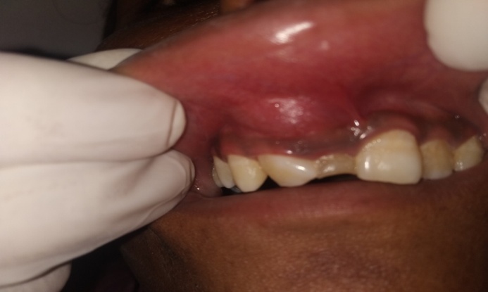
A 15 years old female patient reported in the department of oral medicine and radiology with the chief complain of asymptomatic swelling in upper front right region of jaw since 1 month. Swelling was smaller in size and increased gradually to attain the present size. Medical and dental history was non contributory. Patient was moderately built and nourished and her vital signs were within normal limits. On Extra oral examination diffuse swelling was present on the right middle third region of the face in the region of the nasolabial fold. The skin over the swelling appeared normal and on palpation the swelling was firm in consistency and non tender with the well defined margins. On Intraoral examination a retained tooth 51 which was non vital with missing 11 was seen. A single swelling measuring 1cm to 1.5 cm present in the labial vestibular region extending from labial frenum to the mesial aspect of 13 was seen. The mucosa over the swelling appeared normal in color. On palpation the swelling was non tender and firm in consistency. Correlating all these clinical features a provisional diagnosis of radicular cyst was given and differential diagnosis of dentigerous cyst, adenomatoid odontogenic tumor, calcifying odontogenic cyst was made and the patient was then referred for further radiographic investigations.
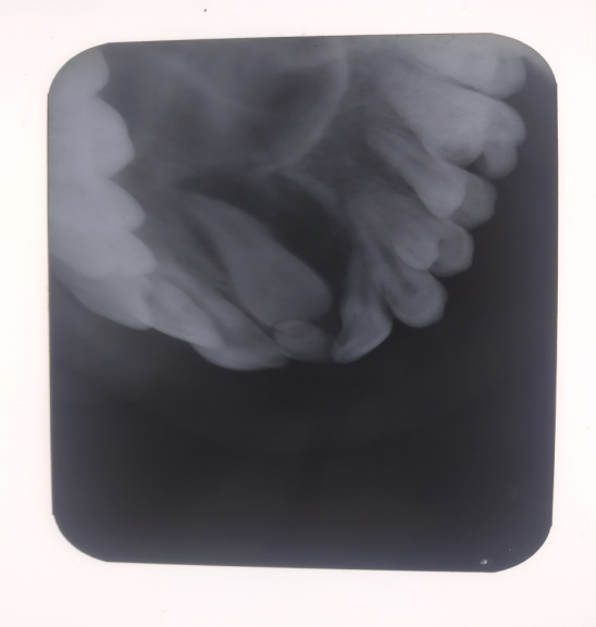
IOPA and maxillary occlusal radiographs (Figure 1, Figure 2) reveals a unilocular radiolucency enclosing the impacted central incisor without any evidence of the calcifications. Incisional biopsy was done under local anesthesia and the specimen was sent for histopathological examination. Histopathological examination gives confirmation of adenomatoid odontogenic tumor. On the basis of above histopathological and radiographical reports final diagnosis of adenomatoid odontogenic tumor was given.
Discussion
AOT was classified into 1st group of tumors in the year 2005 by WHO.4 It is benign, non invasive lesion which three variants namely intraosseous, follicular, peripheral and extrafollicular.7 Mostly the non neoplastic lesions of the oral cavity is the apical cyst, dentigerous cyst, Calcifying Epithelial Odontogenic cyst, Odontogenic Keratocyst, Central Giant Cell Granuloma whereas the common neoplastic lesions are the AOT, unicystic ameloblastoma, ameloblastic fibroma, ameloblastic fibro-odontoma.5 A well demarcated radiolucent lesion associated with the crown of impacted tooth like in case of apical cyst, CEOC, OKC, and CGCG. An apical cyst is mostly associated with the endodontic treated tooth while CEOC, OKC, CGCG are not related to the crown of impacted tooth and are mostly multilocular.5 Dentigerous cyst, unicytic ameloblastoma, ameloblastoma and ameloblastic fibroma are mostly seen in posterior region of mandible and mostly associated with the third molar tooth. While the AOT occurs in the anterior region of maxilla and mostly with the impacted canine tooth.5 The extrafollicular is not associated with impacted tooth whereas the follicular variant is associated with the impacted tooth and the peripheral variant is associated with the gingival structures.7 The origin of AOT is controversial due to which there is a debate still going on whether to keep it in neoplasm or hamartomas. The small size of the tumor and its reccurences in most cases supports that it is a hamartoma on the other hand few authors suggests that its early detection could be the reason for the small size of the lesion whereas the few cases reported its aggressive features gives credibility for the neoplastic origin.8 AOT is an odontogenic epithelial tumor commonly found in female aged between 10-19 years. If geographic aspects are taken in account for gender distribution differences were in Asian and Non-asian races. Asian AOT show a female ratio of 2.3:1. The lesions are asymptomatic but may cause cortical expansion and displacement of the adjacent teeth.7 Maxilla is involved more frequently than mandible. It is commonly associated with the maxillary canine but sometimes it also involves impacted central as well as lateral incisor. Mostly, AOT are central follicular in type and appear as well demarcated radiolucent lesions. AOT surrounds the impacted tooth and it looks like a corticated radiolucency with small radiopacities and if there is no radiopacities is seen then differential diagnosis of dentigerous cyst is given.5 The intraosseous variants appears as a demarcated radiolucencies. Radiolucency contains radioopaque foci. The follicular type mostly mimicks the dentigerous cyst. Aot surrounds the crown of the impacted tooth to laterally extend to the root whereas the extrafollicular type is not associated with the pericoronal relationship and often diagnosed as the lateral periodontal cyst or globumaxillary cyst. The peripheral type may show erosion of the alveolar bone.1 AOT is usually surrounded by well developed connectrive tissue capsule. It may present as a solid mass, large cystic space. The tumor is composed of spindle shaped forming sheets and whorled mases in a connective tissue stroma there is amorphous eosinophilia like material between the epithelial cells as well as in the centre of rosette.2 Dystrophic calcifications are seen in AOTs within the lumen of duct like structures epithelial masses or the stroma.2 The histological typing of WHO defined AOT as a odontogenic tumor in which epithelium contains duct like structures and with the inductive changes in the connective tissue.5 The tumor is well encapsulated therefore, conservative surgical enucleationj is done with the followup of 6 months and the patient was rehabilitated with fixed prosthesis.3
Conclusion
Adenomatoid odontogenic tumor (AOT), due to its slow growing in nature and associated with impacted teeth should always be considered as a possible differential diagnosis of dentigerous cyst. Early diagnosis is important by the clinician as early treatment with conservative surgical enucleation will prevent the destruction of bone, therefore our case presented varied clinical and radiographic manifestations of adenomatoid odontogenic tumor of which a diagnostician should be aware of, while distinguishing more common lesions of odontogenic origin in routine dental examinations.


