Introduction
Maxillomandibular fractures involve a disruption of the tooth, bone, and musculature.1 To achieve a favorable functional outcome, each component must be addressed. Establishing a pre-trauma occlusion for the patient is vital to the treatment plan. Failure to do so results in difficulty in mastication, TMJ dysfunction, periodontal complications, caries-prone teeth, and aesthetic deformities for the patient.2, 3 Since a vast majority of facial fractures are now treated using rigid fixation, maxillomandibular fixation is used only intraoperatively. However, in cases where a non-surgical management of facial fractures is indicated, an MMF is used for a longer period. A myriad of systems for achieving occlusion exists in the literature, for example, arch bars, IMF screws, Ivy eyelet wiring, bonded orthodontic brackets, external pin fixation, embrasure wires, cast metal splints, and pearl steel wires, and acrylic splints.4, 5, 6
Orthodontic brackets have been used for IMF, especially in the pediatric population. However, the bonding strength of the brackets is compromised in the presence of saliva or blood, there are chances of allergic reaction to the resin used and it requires sound dentition for the force exerted by wires.7 The presence of exposed dentin, which is seen in fractured teeth (concomitantly in facial trauma), greatly decreases the bond strength. The fabrication of an acrylic splint requires adept prosthetic skills, specialized equipment, and lab support, and is difficult in patients with concomitant injuries of the body, polytrauma, or admitted in the ICU.8 IMF screws, though proven effective, are not sufficient for use in comminuted fractures. It is also associated with screw loosening and mucosal overgrowth.9
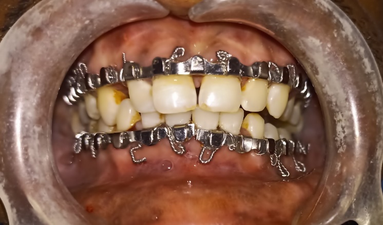
Arch bars are popular because of their tension band effect and stability. However, placing conventional arch bars is cumbersome and time-consuming. There is an inherent risk of wire stick injury, extrusion of teeth due to the traction applied, and unsuitable for patients with periodontally compromised teeth or edentulous dentition.10 There is a need to alleviate the disadvantages of Conventional Erich’s Arch bar (CEAB) and retain the properties of its tension band effect. (Figure 1) Hence CEAB was modified. Perforations were made between the winglets and arch bars were secured using mini-screws in the upper and lower arch. There is a dearth of literature comparing the advantages of modified Screw retained arch bars to the Conventional Erich’s arch bar. Hence, a study was conducted to evaluate the same.
Materials and Methods
A prospective comparative study was conducted on 30 patients with maxillofacial trauma undergoing MMF between the period of April 2022 to October 2022. Ethical clearance was obtained from the Institutional Ethics Committee and the study followed the tenets of the Declaration of Helsinki.
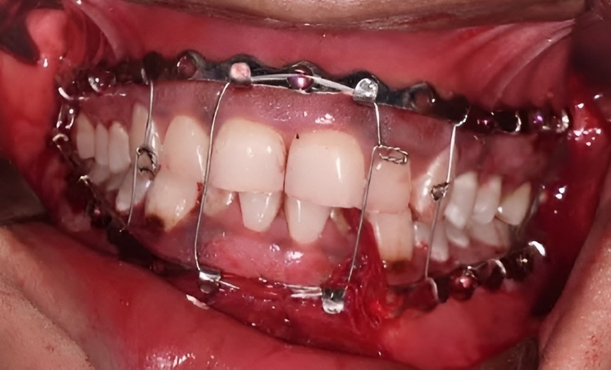
After a thorough pre-surgical evaluation, the patients requiring the application of an arch bar for fracture management were randomly allotted to the test group (Group A) or control group (Group B). Randomization was done using a chit-system. In the test group (Group A) patients received a modified SRAB secured with 1.5 × 10 mm screws which was placed after drilling a suitable-sized hole using a 1.1 mm drill bit in the interradicular and interdental space. (Figure 2) IMF screws were retrieved after the fourth post-operative week in the outpatient department under local anesthesia. The patients in the control group (Group B) received a CEAB secured using 26 gauge stainless steel wire in the standard manner followed by removal after the fourth post-operative week. Patients were evaluated based on the time taken for the application of arch bars, rate of glove perforation and patient compliance during the application, stability of the arch bar, oral hygiene status (using Silness and Loe plaque index), and incidence of complications. Occlusion and mouth opening of the patients were also measured to evaluate the functional outcome. (Table 1)
Table 1
The differences in outcome variables between the two groups differences in outcome variables between the two groups
Exclusion criteria
Medically compromised patients.
Patients with associated bone pathology.
Patients with a previous history of maxillofacial trauma or major reconstructive maxillofacial surgeries.
The data was entered in Microsoft Excel and analyzed using SPSS version 23. The data were subjected to the Shapiro-Wilk test to test the normality. Homogeneity of variance assumption was tested by using Levene statistic homogeneity of variance. The difference in the test variables between the test and control groups was analyzed using the student unpaired t-test and the chi-square test. P<0.05 was considered significant.
Results
A total of 30 patients were included in the study. The time taken in the test group (using modified SRAB) was lower (27.87±5.397) as compared to the control group (using CEAB) (90.20±7.262) and this difference was statistically significant (p=0.000). The rate of glove perforation was lower in the test group (0.20±0.414) as compared to the control group (2.33±1.291) and this difference was statistically significant (p=0.000). In the test group, all patients were compliant as compared to the control group, wherein only 3(20%) of the patients were compliant and this difference was statistically significant (p=0.000). The incidence of complications was higher in the control group (40%) than in the test group (20%). However, this difference was not statistically significant (p=0.232).
At 1 week post-op, the stability of the arch bar was higher in the test group 13(86.7%) as compared to the control group 5(33.3%). At 4 weeks post op all patients in the test group exhibited stability of the archbar whereas only 7(46.7%) of the patients in the control group showed stability of the archbar. The difference between the groups was statistically significant. At 1 week post op the majority of the patients in the test group had excellent oral hygiene status 9(60%) as compared to the control group wherein the majority had a fair oral hygiene status 8(53.3%). At 4 weeks post-op, the majority of the patients in the test group had excellent oral hygiene status 10(66.7%) as compared to the control group wherein the majority had a fair oral hygiene status 10(66.7%).
The incidence of complications was higher in the control group (40%) than in the test group (20%). At evaluation 2 (week 1), the incidence of complications was the same in both groups. At evaluation 3 (week 4), the incidence was complications was slightly higher in the control group 4(26.7%) as compared to the test group 3(20%). They were not statistically significant.
The mean mouth opening in the test group was 38.80±3.342 and in the control group, it was 38.73±3.327and this difference was not statistically significant. At evaluation 3 (4 weeks post-op), 13 (86.7%) patients in the test group had normal occlusion, and 8 (53.3%) patients in the control group had normal occlusion, and this difference was statistically different (p=0.046).
Discussion
Attaining a stable, pre-trauma occlusion is one of the tenets of treating facial fractures. Intermaxillary fixation methods have evolved through the years, to optimize the treatment time, minimize patient discomfort, and reduce risk to the operator. At the same time, the functional outcome of fracture management should not be compromised. The tension band effect an arch bar exerts is its most redeeming quality. The conventional Erich’s arch bar used stainless steel wires, and a sound dentition that was not periodontally compromised was essential for its application. Hybrid arch bars were a choice alternative and were secured to the maxillary and mandibular arches using mini-screws. However, they had certain drawbacks like mucosal coverage of screws and positioning. A novel modification of CEAB done by Queiroz et al. in 2013, was done by drilling screws in the arch bar.11 Our study compares the CEAB with the modified SRAB on various parameters to help guide clinicians for a superior treatment outcome.
Based on our study results the mean time taken for the application of a modified SRAB (27.87±5.397) was lesser as compared to CEAB (90.20±7.262). Duration of operating time is an independent factor in determining post-operative complications. Surgeons across the specialties should strive to reduce operating time.12 This study recorded the rate of glove perforation to be lesser in the modified SRAB group (0.20±0.414) as compared to the CEAB group (2.33±1.291). Maxillofacial trauma surgeons deal with a huge number of patients with blood-borne diseases.13 Needle stick injury and wire-prick injury can lead to adverse effects for the entire operating team. It is imperative to adopt treatment modalities that reduce this risk. A study conducted by Pathak et al. reported similar results concerning time taken and glove perforation.14
In the current study, all patients were compliant during the application of the arch bar in the modified SRAB group as compared to the CEAB group, wherein only 3(20%) of the patients were compliant. The results also reveal greater stability of the modified SRAB compared to the CEAB at both evaluations. This is in agreement with data from another study by V. Venugopalan et al. comparing modified Erich’s arch bar with CEAB.15 The present study revealed a lesser incidence of complications with modified SRAB (26.66%) as compared to CEAB (35.55%). Accidental tooth root injury during pre-drilling for screw placement is a major complication of SRAB. In case of injury to the root pulp, endodontic therapy is required to retain the tooth in the mouth. A study conducted by Hartwig et al. reported that despite frequent iatrogenic tooth root injury, the adverse effects were rare.16 The authors also recommend the placement of screws in safe zones around the tooth to avoid such injuries. Drill breakage was another complication noted in the present study. Retrieval of broken drill bits necessitates the removal of surrounding bone, increases the operating time, and offers scope for further complications.17 The study recorded significantly better oral hygiene in patients with modified SRAB than CEAB. This is in line with previous studies comparing conducted by Pathak et al.14 Poor oral hygiene can have detrimental effects on intraoral wound healing and is associated with a risk of surgical site infection.18, 19 However, the results of this study differ from those of Hamid et al. who reported no significant difference in oral hygiene (measured by GI scores) between screw-retained and Erich’s arch bar.20
Whichever modality of achieving MMF is chosen, the treatment of facial fractures should not be compromised. This study was unique in measuring the functional outcomes between the two types of arch bars at the end of 4 weeks. The results show that the difference between the test and control groups was not statistically significant and the patients in both groups had acceptable occlusion.
Conclusion
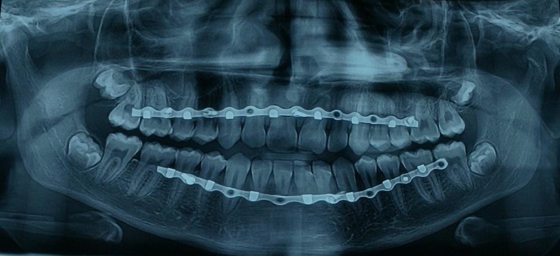
Based on the results of the study we can conclude that the modified screw retained arch bar is a better alternative to the conventional Erich’s arch bar in MMF for maxillofacial fractures. The use of modified SRAB has a shallow learning curve. It can be easily taught to residents and clinicians. However, the authors advise using an orthopantomogram to identify the position and anatomical variations in roots. (Figure 3) This will reduce the number of inadvertent root injuries. One of the main disadvantages of SRAB is that the position of the screw holes needs to coincide with the inter-radicular space. A proper pre-operative analysis will help in this aspect. The position of the modified arch bar might interfere with the placement of incisions in the symphyseal or parasymphyseal region. The authors advise modifying the mucosal incision to a lower level, and removal of the arch bar after fracture fixation for proper closure.21
Ethics Statement/Confirmation of Patients’ Permission
Ethical clearance was obtained by the Institutional Ethics Committee at SGITO, Bengaluru. All patients gave written consent to their inclusion in the study.


